THE SHROUD
THEORIES and EVIDENCE
Recent discoveries have shed new light on the unique and unexplainable aspects of the shroud. In other words, now more that ever we're forced to accept that the shroud is something beyond human ability to produce; even with our highly advanced technologies. This fact, coupled with the data contained in the image, puts us in a very interesting yes or no dilemma. All the data describes Jesus. We're faced with accepting the God of the Bible or rejecting much irrefutable evidence. The only explanation for the shroud is Jesus and his resurrection. How can we accept that someone came back to life after 72 hours? But how can we deny our own senses and intelligence?
"Resurrected" is the name of the book I'll be sharing with you on this page. It was written by Gilbert R. Lavoie, M.D. and published in 2000 by Thomas More. It contains some important updating of old information, and some brand new aspects of the old evidence.
In the introduction, Gary Habermas, Ph.D, D.D., says:
Let me warn you that the author is a man of science. Dr. Lavoie was convinced that the shroud was the real thing until 1988 when he heard the news of the carbon dating results. He did not take the view that the shroud was authentic at all costs. Rather, he assumed that the cloth was a fake, and told his audiences so. But later he changed his mind, convinced that the accumulation of scientific studies more than overwhelmed the carbon dating results. ñGary Habermas, Ph.D., D.D.
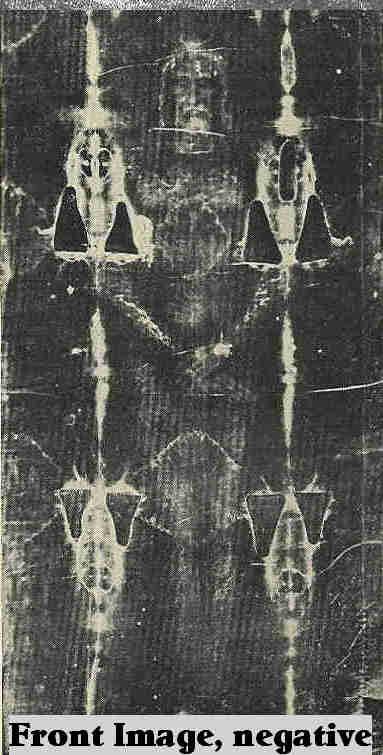
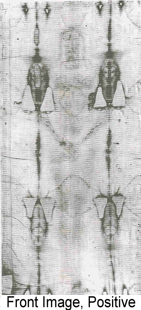
A French surgeon, Dr. Pierre Barbet wrote: A Doctor at Calvary. His work centered on a cloth that displayed the blood-marked image of a naked man. This linen cloth, approximately three and a half by fourteen feet, is known as the Shroud of Turin.
Dr. Barbet had his first direct experience with the shroud during the Turin exposition in 1933. He was on the steps of the cathedral, and with the help of the light of day, he saw the following:
From a distance of less than a yard and I suddenly experienced one of the most powerful emotions of my life. For, without expecting it, I saw that all the images of the wounds were of a color quite different from that of the rest of the body; and this color was that of dried blood which had sunk into the stuff There was thus more than the brown stains on the shroud reproducing the outline of a corpse.
The blood itself had colored the stuff by direct contact, and that is why the images of the wounds are positive while all of the rest is negative.
After reading this, I looked again at the photographs, and it was true. The blood was positive on the cloth while the image was negative. In contrast, the blood was negative on the photographic negative of the cloth while the image was positive. This meant that the blood marks were photographically opposite to the image that they accompanied. What pictures carry with them photographic opposites? Other than the shroud, I knew of none.
According to Barbet's eyewitness report, the blood marks were encrusted and intertwined with the fibers of the cloth. As per Barbet, "The blood itself had colored the stuff by direct contact.î It was his experience with wounds and bandages as a surgeon that led him to conclude that the blood marks on the shroud were not what people had been casually calling blood flows, but were actual imprints of blood clots on cloth.
In contrast to the blood marks, the fibers of the image of the man were devoid of anything but the brownish fibers themselves. All that I knew was that he had not seen paint, but only the ìbrown stains" that made up the image. I did not understand what those brown stains were, but neither did he.
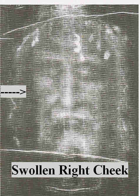 His observations of the face, especially the swelling under the right eye, caused me to stop and pay closer attention to the details. At first I did not see the swelling, but after comparing the area under the left eye with that of the right, I could see what Barbet was referring to. It was as if the man had been struck in the face with a fist or a stick at the area of the cheekbone.
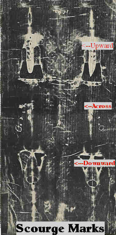 Just as revealing as his observations of the face was his study of the scourge marks. These wounds are all over the body but are best seen on the back image. The stripes on the back image are oblique, slanting upward toward the right and left shoulders. On the buttocks, they change position and are no longer oblique; they are horizontal. On the legs, they are again oblique, slanting downward toward the left.
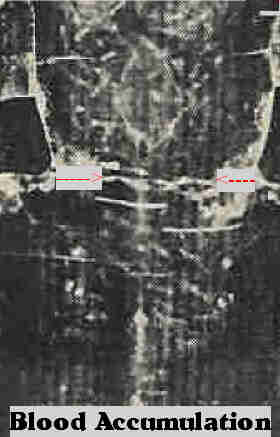 Barbet studied all the major blood marks: the cap of thorns, the side wound, the blood marks of the feet, and the horizontal blood flow at the small of the back. This flow occurred after the man was taken from the vertical position of crucifixion and placed in the horizontal position in preparation for burial.
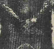 One thing puzzled Barbet. The image of the hands showed no thumbs! On the shroud, the two hands when seen from behind only show four fingers with the thumbs hidden in the palms. Barbet himself did not know the reason for hands without thumbs until he plunged a nail through the wrist of a freshly amputated arm.
When the nail went through the soft anterior parts of the wrist, the palm being upwards, the thumb would bend sharply and would be exactly facing the palm by the contraction of the thenar muscles, while the four fingers bent very slightly.
How could a 14thñcentury artist have been so anatomically precise, and moreover, why would he attempt to be so precise. Was the artist anticipating the scrutiny of a 20th-century surgeon? Furthermore, this artist would have had to create a negative image five hundred years before the invention of the camera. Then he would have had to put the image on cloth without the use of paint.
Barbet had presented convincing medical evidence demonstrating that this cloth once held the body of a man that had been both scourged and crucified.
FLOWERS IN THE TOMB
The shroud has been newly investigated several times. Each time a serious threat of fraud has been raised; a new look has surpassed refutation of the shroudís authenticity. First, ìIt was painted.î Next, ìC14 dating says itís new.î
Dr. Max Frei, was a professor of criminology from the University of Zurich. He was a biologist who made a specialty of using microscopic techniques in the field of criminology. He had discovered pollen grains on the shroud and thus was able to give it a geographical history.
ìA positive identification of such pollens would be a confirmation of the shroudís stay I a particular botanical region. From 1974 to 1978 I raveled several times (in different floreal seasons) through Palestine, Turkey, through Cyprus, France and Italy, collecting pollens for direct comparison under the microscope. I succeeded in identifying 57 different plats which have left microscopical evidence on the shroud.
None of the pollens was glued to the cloth with tempera or covered with tempera. This is strong evidence against the possibility of the shroudís being a painted fake.
Plants on the Shroud from Palestine and Anatolia Are so numerous compared to the species from Europe, that a casual contamination or a pollen transport from the Near East by storms in different seasons cannot be responsible for their presence, as I have explained in several conferences and publications. The predominance of these pollens must be the result of the Shroud's stay in such countries where these plants form part of the normal vegetation.
Frei leaves us with two very interesting pieces of information: (1) Even though the known history of the shroud is confined to France and Italy from the 1350s on, with Frei's pollen study we now know that the shroud did have an earlier Asian and Holyland history that preceded the 1350s. This history was either unknown or for some reason never passed on by its first recorded European owner, Geoffrey de Charny. (2) Frei found no tempera on the pollens of the shroud. Frei felt that this was strong evidence against the shroud image being a painting.
A new dimension has been added to Freiís study through the work of Alan Whanger, M.D., and his wife Mary. I 1989, while attending a shroud symposium in Paris, the Whangers showed me their observations of very faint images of flora on the non-image areas of the shroud, mainly about the head of the image. They identified 28 species of plants, all found in Israel.
In 1995 the Whangers went to Israel seeking the help of Professor Avinoam Danin, and authority on the plant life of Israel, who works at the Alexander Silberman Institute of the Life Sciences at the Hebrew University, which published an article saying:
Danin examined their [Whangers'} findings, verified them, and even determined that additional images found on the garment could be associated with plants from the land of Israel. Danin found images of summer and winter leaves of zygophyllum which are characteristic of the plant pollen grains from the rockrose [which} were recovered from the shroud in the 1970s by Dr. Max Frei, a forensic scientific expert from Switzerland. This kind of "double proof" (images and pollen) shows conclusively that there were indeed rockroses that were placed with the shroud, says Prof. Danin.
Prof. Danin says that his research shows that the types of plants found on the shroud-when compared to his mapping of these same plants as found in nature-show that 70 percent of these species can be found within a 10x10 square kilometer area whose center lies in an area between
Jerusalem and Jericho. Prof. Danin noted further that zygophyllum grows only in Israel, Jordan and Sinai, with its northernmost boundary in the world being at the sea level sign on the highway between Jerusalem and Jericho. In view of this, one can narrow down even further the origin of the shroud and say definitely that one is dealing with the Jerusalem area. The fact that a winter leaf was found on the zygophyllum, together with remnants of the stalk from the preceding year, proves, say Prof. Danin, that it was plucked in the spring, which was the season identified with most of the plants revealed on the shroud.
The Whangers and Professor Danin thus complement Frei's work and present evidence that the shroud originated in the Jerusalem area in the spring of the year which is the time of the Passover. A precedence for using flowers in Jewish burials is found in 2 Chronicles 16:14 (verbal communication with Anthony Opisso, M.D.)
THE IMAGE OF EDESSA
Frei's itinerary of the shroud, as determined by the pollens, also coincided with Ian Wilson's theories as to the whereabouts of the shroud prior to the 1350s. Ian Wilson, a history graduate of Oxford and a journalist, is the author of the book The Shroud of Turin, published in 1978. He was also presenting at the conference, which gave me the opportunity to hear about his work firsthand. His book is a compilation of fragmented historical reports that tell of an image-bearing linen cloth that carried the likeness of Jesus. He begins his story in the early part of the first century and takes us from Jerusalem to the ancient city of Edessa, now called Urfa, located in the southeastern part of Turkey. There, an image-bearing cloth was given to the incumbent king, Abgar, where it remained hidden for five centuries. It resurfaced at the same site in A.D. 544 as "the divinely made image not made by the hands of man" as it was reported by the Syrian historian Evagrius (527-600). Regarding the image, Wilson quotes Edward Gibbon's The Decline and Fall of the Roman Empire:
Before the end of the sixth century these images made without hands. . . were propagated in the camps and cities of the Eastern empire; they were the objects of worship and the instruments of
miracles; and in the hour of danger or tumult their venerable presence could revive the hope, rekindle the courage, or repress the fury of the Roman legions.
In A.D. 944, the image of Edessa, also known as the Mandylion, which Wilson believes to be the shroud, arrived in Constantinople where it remained for over 250 years. Wilson's theory is interesting, but he has to contend with the fact that the Mandylion was thought to have been a facial image whereas the shroud displays a total body image. In 1998, Wilson documents new evidence to back his theory. He writes:
In a 944 AD sermon, Gregory, archdeacon of Hagia Sophia, Constantinople, delivers a sermon in which he imparts that it bears not only "'the drops of sweat from the agony [in Gethsemane] which flowed from [Christ's] face like drops of blood but also blood and water [haima kai hudor] from his very side. " This indicates (1), that there was more to the imprint on the cloth of Edessa than just a face; and (2), that the imprint included blood of the Passion and must thereby have been created after Jesus was taken down from the cross, i.e.,. precisely corresponding to the Turin Shroud.
Wilson's most convincing documentation locates the shroud in Constantinople. From the pages of the diary of a French crusader by the name of Robert de Clari, we find the following account:
"There was another of the churches which they called My Lady St. Mary of Blachernae, where was kept the sydoine [shroud cloth] in which Our Lord had been wrapped, which stood up straight every Friday so that the figure of Our Lord could be plainly seen there. And no one, either Greek or French, ever knew what became of this sydoine after the city was taken." This was written during the first part of the 1200s after Constantinople was sacked by the army of the Fourth Crusade.
X-RAYS
Dr. John Jackson, a physicist, placed a photograph of the shroud image under a VP8 image analyzer. This electronic device is ìÖideally suited for determining whether a given image contains distance information because it converts image shading into relief.î Jackson found that the frontal image of the shroud has a three-dimensional quality. Photographs of people do not exhibit this quality. Much later, in 1992, Whanger discovered another quality about the image. He found ìX-ray-like images of internal body structures? Such as bones of the face and hands.
This is a unique and complex image that one may say is more than a photograph: it seems to possess some characteristics of an X-ray, and carries with it information that gives it a three-dimensional quality.
THE MICROGRAPHS
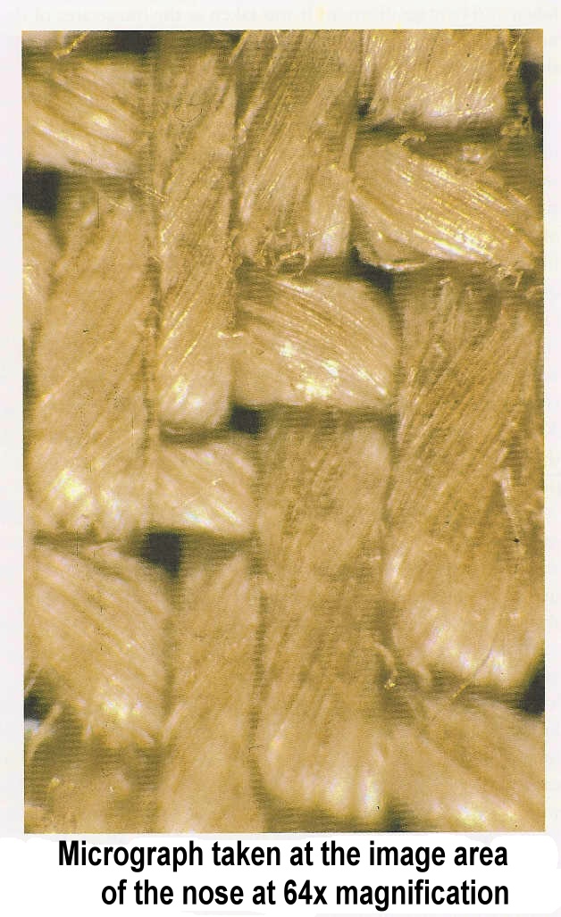
Vern Miller, one of the official professional photographers for the American scientific team has a micrograph (a slide) that was taken at 64x magnification. It was taken at the image area of the nose, which happens to be the darkest portion of the entire shroud image.
The individual fibers of each thread are yellowed. It is these individual yellowed fibers, not paint, that cause the image.
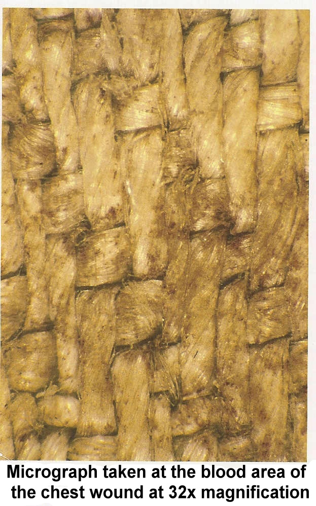
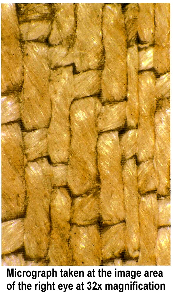
Another micrograph, taken at the blood area of the chest wound at 32x magnification, showed that in and about the fibers is intertwined debris that Barbet had believed to be blood. A third micrograph was taken at the eye area at 32x magnification. In the eye and the nose micrographs of the image, NO DEBRIS can be seen.
CHEMICAL ANALYSIS
Two questions needed answering: (1) what caused the yellowing of the fibers that caused the image and (2) what was the debris that Barbet believed to be blood.
Sticking tapes that had been placed over the blood marks and image marks of the shroud were given to a man by the name of Dr. Alan Adler. Adler, a professor of chemistry from western Connecticut State University, is an expert on porphyrins, which are organic chemicals that form part of the structure of red blood cells. Adler analyzed the particles and fibers that adhered to the sticking-tapes. He eventually ìestablished by detection of heme derivatives, bile pigments, and proteins,î the presence of whole blood on the shroud.
SHINING THOUGH
Adler says, ìYou know, one of the best tests they did during those five days of testing in Turin was one of the simplest tests they did.î They shone a light through the cloth. As the light passed through the cloth, the blood images could be easily seen, but the body image could not be seen. What did this mean? It meant that the image was not made from an applied substance, but was contained in the very fibers of each thread. NO PAINT! The scientific team also discovered that the blood marks had soaked through to the backside of the shroud, whereas the image marks were not seen from the back of the cloth. But more astounding proof was to come out of the micrographs.
TEENY TINY FIBERS
Adler found that a single thread of the shroud cloth was made up of many very small (10 to 15 microns in diameter) linen fibers. The width of one of these fibers is much smaller than a human hair. Only the TOPMOST fibers of the thread were yellowed. These yellowed fibers, composed of cellulose, were found only on the image side of the cloth and were responsible for the image. The fibers on the bottom side of the threads were NOT yellowed.
Adler concluded that the yellowing of the shroud image fibers was produced by a dehydrative, oxidative process that affected the fiber (cellulose) structure itself and caused it to yellow. In other words, this process is a degradation of the fibers themselves and is identical to the aging of linen, causing linen to turn from white to yellow.
WHICH CAME FIRST?
On removing the blood from the blood-covered fibers, Adler found that these fibers, instead of being yellow as they were at the image areas, were white. That meant that (1) the blood went onto the cloth before the image marks and (2) that the blood protected the fibers from whatever caused the image. Therefore, the blood came first; the image came later.
SOME ARE, SOME ARENíT
This next part is truly amazing and confirms absolutely the supernatural quality of the image. Remember those Teeny Tiny Fibers?
Dr. Eric Jumper, Notre Dame professor found that as the yellowed uppermost fibers dipped down under another thread, they were no longer yellow but remained their original white. Whatever caused the image affected only the uppermost fibers, even to the point of not wicking along the fiber as a liquid would do if it had been applied to the shroud surface.
WHATEVER CAUSED THE YELLOWING OF THE SHROUD FIBERS PENETRATED DOWN INTO EACH THREAD THE DEPTH OF ONLY ON FIBER!!! NOT AS DEEP AS A HUMAN HAIR!!!
But thereís more. All those top fibers turning yellow would have made a ìwashî of yellowing, not an image of many different shades. How then was it possible that we see an image? Itís just like the pictures in the newspaper.
Jumper saw that there were examples everywhere of yellowed image fibers lying side by side to white non-image fibers. He also noticed that the yellowing of the individual fibers was uniform; each fiber held almost exactly the same quantity of yellowness.
It turned out that the difference in the shading of yellow from one area of the image to another was dependent on the NUMBER OF YELLOWED FIBERS PRESENT! ìItís like the dots of newspaper print. If you want to make an area darker, you put in more dots.î
Dr. Adler pinned it all down by saying, ìFor a painter to have created this image, he would have needed a paintbrush the size of a fiber which is less than half the width of a human hair.î
A paintbrush the size of one pixel? I don't think so.
PEOPLE WILL SAY ANYTHING!
The New York Times carried three articles on October 14, 1988. The first summed it up:
ìRadiocarbon tests conducted independently by three laboratories this yearÖ.concluded that the shroud cloth was created between 1260 and 1390.î
It turns out that the testing wasn't even done by the scientist's own protocol. They wanted six samples of the shroud and the use of seven laboratories in order to assure the best result. The reality is that the protocol was abandoned and the scientists, through no fault of their own, were given only one sample of the shroud. This sample was portioned and three pieces were the given to three radiocarbon dating labs, each lab receiving on piece of this one sample.
Dr. Alan Adler examined a piece of the sample, comparing it to the non-image area by Courier Transform Infrared microspectrophotometry and by a scanning electron microprobe. The results clearly indicate differences in chemical composition. îThe radiocarbon samples are not representative of the non-image sample that comprise the bulk of the cloth." Therefore, the sample given to the radiocarbon scientists does not follow their own criteria that a sample be ìrepresentativeî and ìhomogeneous.î
As amazing as the above evidence is, there is still more. There is a need to explain how: the cloth could be tucked down around the body, but the image remain flat; that the image is not of a man laying down, but in an upright posture, and yet, not standing; that there are shadows that show the "lighting of the image" came from above the head.

I love mail. 
Come Home